Other studies / 03
The immune microenvironment in humanized models
Studied mast-cell interactions with the immune microenvironment in humanized mouse models. Combined immunofluorescence, flow cytometry, magnetic sorting and 10x Genomics with timed interventions and tumor-growth observations.
- Period
- 2023.09—2024.02
- Research focus
- A*STAR · Mast cells · Breast cancer
Research overview
This work uses humanized mouse models to investigate interactions between breast cancer cells and the immune microenvironment, with a focus on mast cells. The methods bring together tissue observation, cell sorting and single-cell technologies.
Functional experiments selectively inhibited mast-cell activity at different time points and assessed subsequent tumor growth rates. The aim was to explore mast cells’ role in breast cancer progression and their potential as a therapeutic target within this model.
Figures
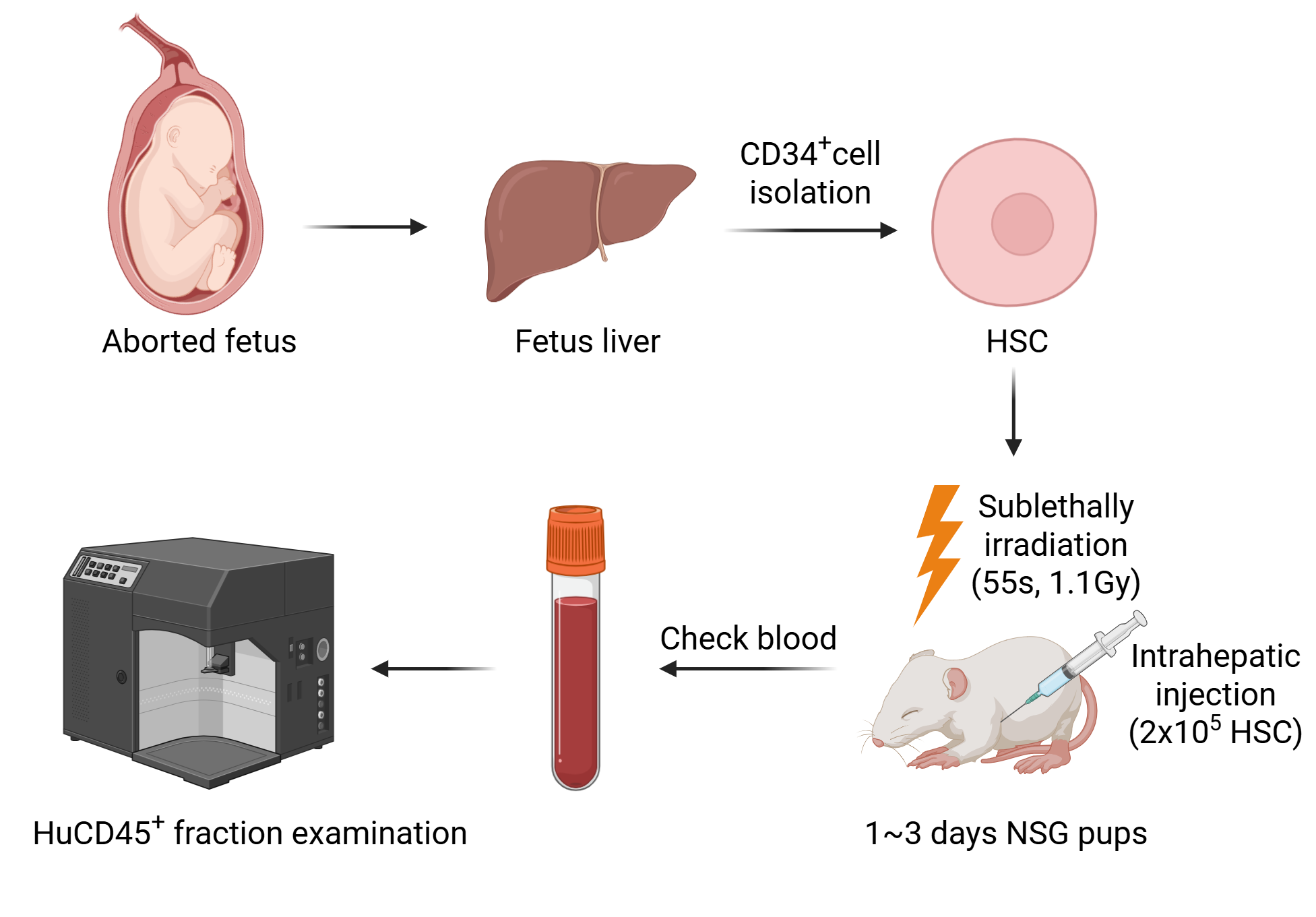
Establishing and checking a humanized immune model
The sequence establishes the humanized-model context before discussing tumor growth and immune composition. The workflow shows engraftment and peripheral-blood assessment, defining the experimental system for the intervention comparisons that follow.
The route runs from donor-cell material through host preparation and engraftment to peripheral-blood assessment. It establishes the model context for later tumor and immune-intervention observations.
Takeaway
Model characterization defines the system producing the later results, not treatment efficacy. The next page compares tumor growth under individual and combined interventions.
Tumor-growth trajectories under single and combined interventions
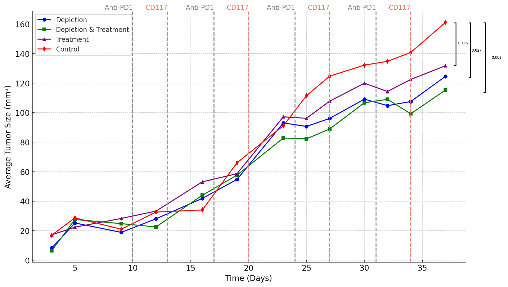
This page compares tumor-growth courses across intervention combinations. Locate the Anti-PD1 and CD117 timing markers, then examine when trajectories diverge and which groups the endpoint brackets compare, rather than only ranking final tumor sizes.
Time is plotted against average tumor size for Depletion, Depletion & Treatment, Treatment and Control. Dashed Anti-PD1 and CD117 markers identify intervention times, and right-hand brackets show the original endpoint comparisons.
Takeaway
The curves describe the overall tumor phenotype without identifying which immune population drives it. The next two pages add cell-composition observations at different time points.
Day 20: immune-cell composition across samples
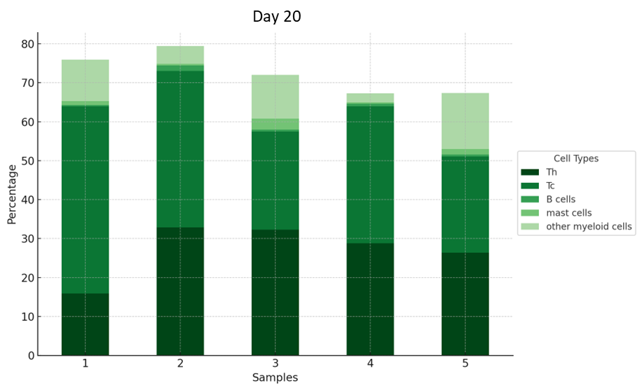
The focus shifts from the overall tumor phenotype to day-20 cell composition. Each bar represents a sample and colors identify cell types; compare proportions while retaining sample identity rather than assigning unprovided treatment labels to sample numbers.
Stacked bars for samples 1–5 display Th, Tc, B cells, mast cells and other myeloid cells. Read both between-sample variation and within-sample composition; sample numbers are not automatically treatment labels.
Takeaway
The proportions describe the cellular environment at this time point, not necessarily changes in absolute cell counts. The next page adds day 31 to the temporal picture.
Day 31: later composition and the pooled sample
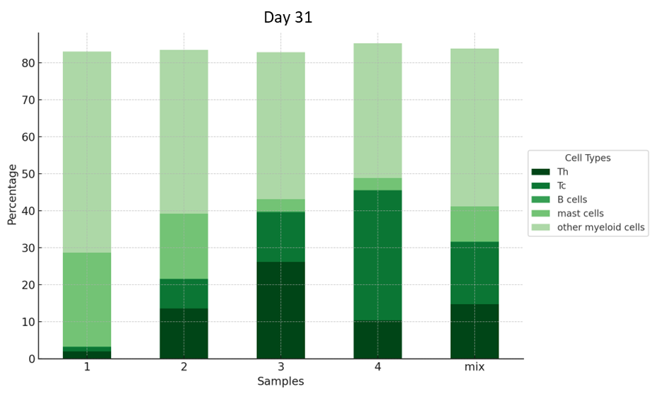
The final page adds day-31 composition using the preceding cell-type colors. Retain the distinction between numbered and mixed samples: a pooled sample is not an additional independent individual, and matching numbers should not automatically be assumed to be paired across days.
Day 31 uses the same cell-type legend for samples 1–4 and a mix. It adds a later time point while retaining the distinction between individual and pooled samples.
Takeaway
The sequence connects model establishment, intervention timing, tumor growth and immune composition. Descriptive differences across time motivate further analysis but do not alone establish a causal link from a cell-population change to treatment response.
My contribution
I conducted orthotopic breast cancer studies in humanized mice, combining frozen sections, immunofluorescence, flow cytometry, magnetic sorting and 10x Genomics. Functional experiments included mast-cell interventions, combinations with anti-PD-1 and tumor-growth observations, connecting cell and tissue analysis with in vivo studies.
Outcomes & record
This work formed part of my visiting-student appointment at A*STAR in Singapore from September 2023 to February 2024. It provided experimental experience combining tissue observation, cell sorting and functional intervention to study the tumor immune microenvironment in humanized mouse models.