Other studies / 02
Circulating tumor cells across cancers
Established DNA-barcode delivery protocols, built CDX models and isolated CTCs from clinical blood samples. Contributed to SMART-seq3 and 10x Genomics RNA libraries for over 1,000 individual cells, with further experimental work on fatty acid oxidation.
- Period
- 2021.06—
- Research focus
- DNA barcoding · Single-cell libraries · Metabolism
Research overview
This work combines gene-library construction and pan-cancer investigation of circulating tumor cells (CTCs), spanning cell-line barcoding, isolation from clinical blood samples, single-cell RNA library preparation and tumor observation in animal models.
Following leads from single-cell RNA sequencing and spatial metabolomics, the experimental work focused on fatty acid oxidation (FAO) in melanoma brain metastasis and the relationship between L-carnitine, CPT1A, SREBP2 / mevalonate (MVA) signaling and cholesterol metabolism.
Figures
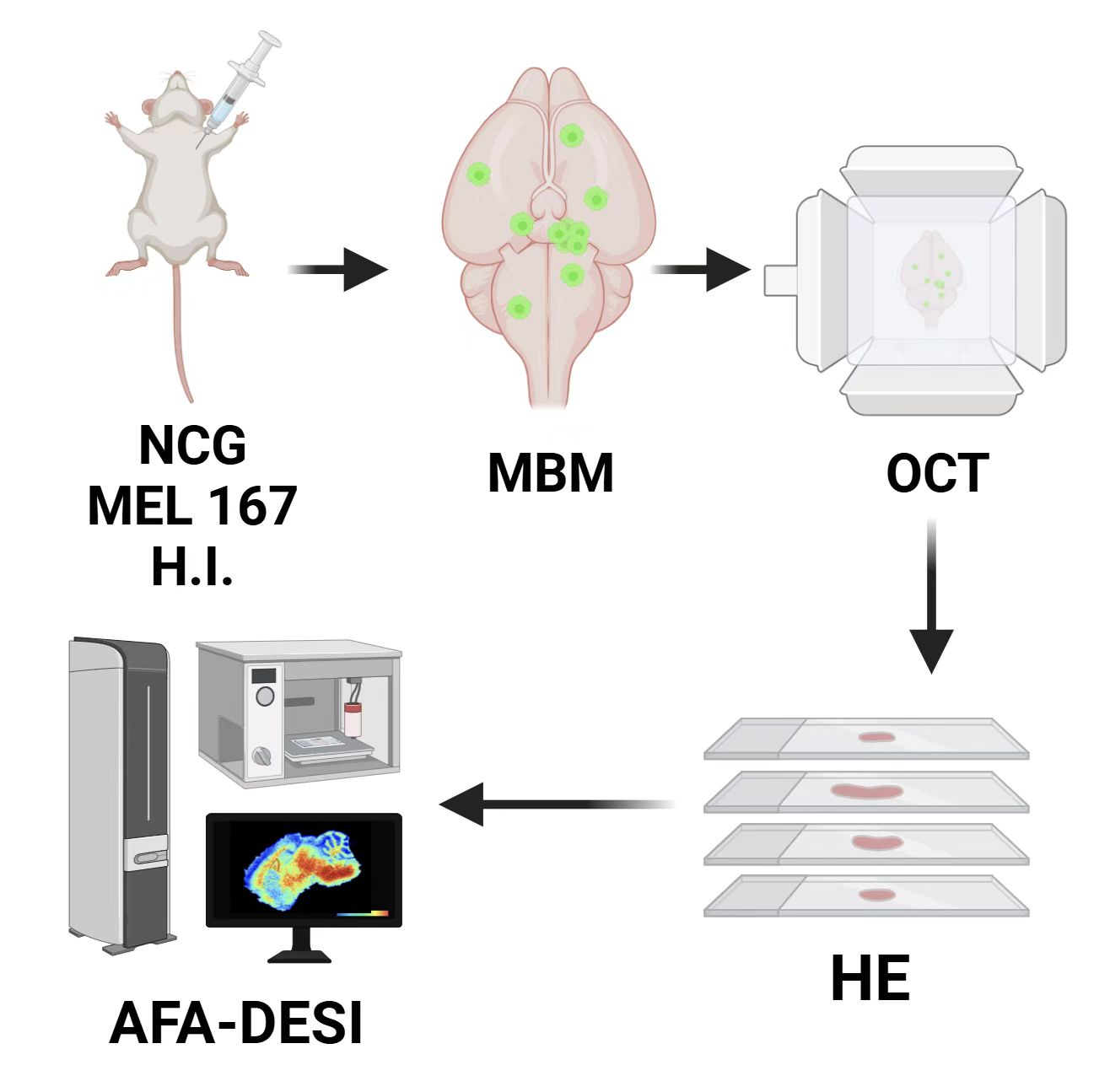
Connecting a melanoma brain-metastasis model to spatial metabolomics
The sequence first establishes how material was generated for the brain-metastasis and fatty-acid-oxidation subproject. The model, tissue preparation and spatial analysis define the context for the subsequent imaging and intervention figures.
The arrows connect the NCG/MEL 167 model to brain-metastasis tissue, OCT embedding, sections and AFA-DESI spatial metabolomics. This is the brain-metastasis/FAO subproject within the broader CTC research experience, not the DNA-barcoding procedure itself.
Takeaway
This page explains how material reaches the analysis; the next asks where the metabolic signal occurs within tissue. Model preparation and downstream spatial-metabolomics work remain distinct steps.
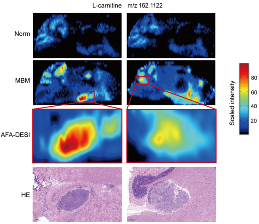
Spatial L-carnitine signal in brain-metastasis regions
With the model and preparation defined, this page examines spatial correspondence between the L-carnitine-associated signal and lesions. Locate the signal in the tissue first, then compare enlarged regions with H&E rather than interpreting a high-signal patch alone.
Rows show Norm, MBM, enlarged AFA-DESI regions and H&E from top to bottom, using the displayed scale for L-carnitine-associated signal at m/z 162.1122. The question is spatial correspondence between molecular signal and lesion location.
Takeaway
Spatial correspondence motivates functional testing rather than directly proving that the metabolite drives lesion formation. The next page introduces L-carnitine treatment and follows the in vivo signal over time.
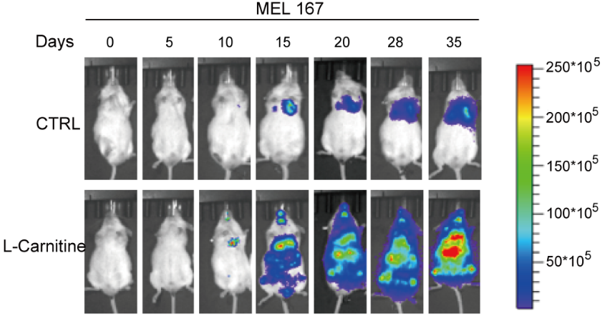
Longitudinal tumor-associated signal with L-carnitine supplementation
Following the spatial-metabolic observation, L-carnitine treatment examines its relationship to in vivo progression. Read both the within-group time course and between-group comparisons at matched dates, retaining the displayed imaging scale.
Rows compare CTRL and L-Carnitine across days 0, 5, 10, 15, 20, 28 and 35. Follow progression within each row before comparing conditions at matched time points rather than selecting only the final image.
Takeaway
The longitudinal images provide a post-intervention readout, not a complete mechanism. The next page examines genetic and pharmacologic CPT1A perturbations as another functional comparison within the project.
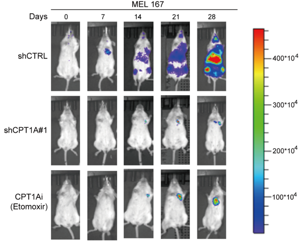
Genetic and pharmacological tests of a CPT1A-associated pathway
This page approaches the pathway differently from L-carnitine supplementation, using CPT1A depletion and inhibition. Follow each row over time, then compare groups at matched dates to relate progression to the different perturbations.
The three rows compare shCTRL, shCPT1A#1 and CPT1Ai (Etomoxir) on days 0, 7, 14, 21 and 28. Unlike L-carnitine supplementation, these interventions probe the pathway by reducing CPT1A-associated activity through genetic and pharmacological approaches.
Takeaway
The four pages move from model and sampling to spatial clues, metabolite intervention and pathway perturbation. Imaging trends remain tied to group quantification and experimental context; they do not establish organ-specific effects absent from the displayed evidence.
My contribution
My work included establishing barcode-delivery protocols, generating cell lines, constructing CDX models, performing in vivo imaging and isolating clinical CTCs. I also contributed to RNA library preparation for over 1,000 individual cells and performed FAO perturbation and cholesterol-rescue experiments.
For the spatial-metabolomics work, I constructed animal models and prepared tissue sections; the collaborating team carried out spatial-metabolomics analysis.
Outcomes & record
My related publications include a first-author paper in BMJ Oncology on CTC-cluster isolation, biological significance and therapeutic implications, and a coauthored paper in Aging and Disease on a potential “anti-Warburg effect” in CTC-mediated metastatic progression. Both were published in 2024.