Biomed / 01
Circulating tumor cell clusters
A review of the isolation, biological significance and therapeutic implications of circulating tumor cell clusters. Published in BMJ Oncology; I am the first author.
- Publication status
- Published
- Journal
- BMJ Oncology
- Authorship
Abstract
Circulating tumour cells (CTCs) and CTC clusters are considered metastatic precursors due to their ability to seed distant metastasis. However, navigating the bloodstream presents a significant challenge for CTCs, as they must endure fluid shear forces and resist detachment-induced anoikis. Consequently, while a large number of cells from the primary tumour may enter the circulation, only a tiny fraction will result in metastasis.
Nevertheless, the metastatic potency dramatically increases when CTCs travel in conjunction with other cell types to form CTC clusters, including neutrophils, myeloid-derived suppressor cells, macrophages, platelets, cancer-associated fibroblasts and red blood cells found in circulation. Such heterotypic CTC clustering events have been identified in a variety of cancer types and may serve as intriguing therapeutic targets and novel biomarkers for liquid biopsy.
This review summarises recent advances in microfluidic technologies designed for the isolation of CTC clusters and explores the biological properties of distinct types of CTC clusters within the circulatory system. Investigation of the mechanisms of CTC cluster–blood microenvironment interactions may offer a promising avenue for gaining fresh insights into CTC cluster-mediated metastatic progression and reveal potential opportunities for devising personalised antimetastasis treatments.
Read paper ↗Figures
Microfluidic isolation of CTC clusters
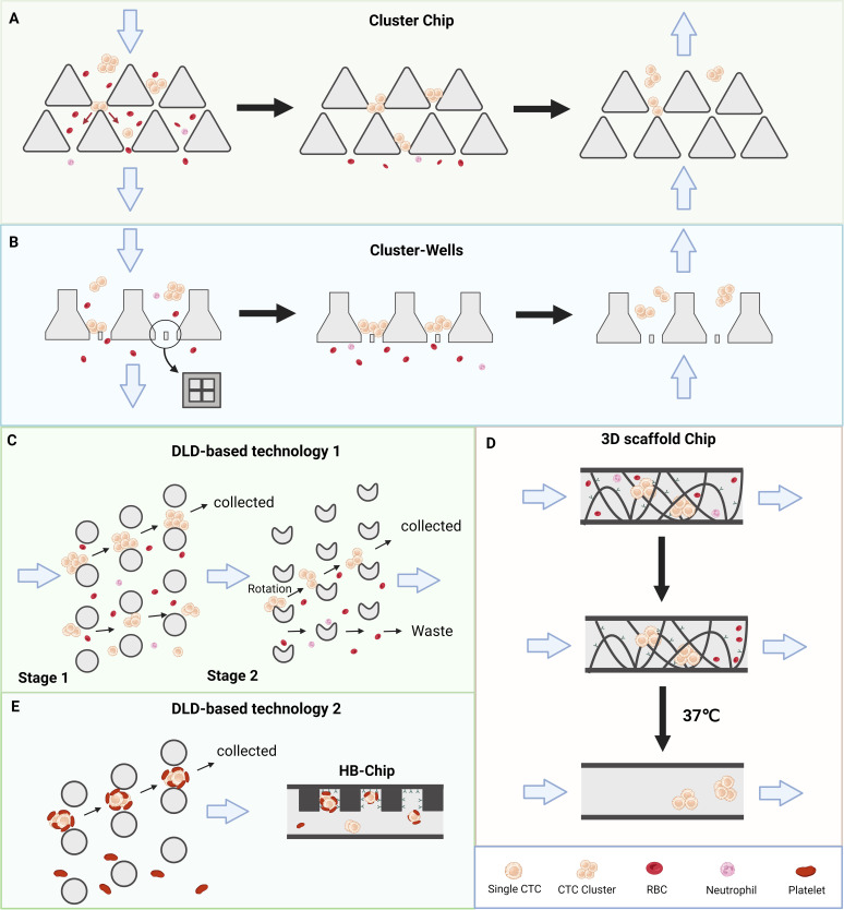
Studying CTC clusters first requires recovering them intact from a vast excess of blood cells. Clusters differ in size and may contain partners such as platelets; breaking them apart during isolation would erase the interactions being investigated. Figure 1 follows the engineering choices from physical filtration and trajectory-based sorting to releasable affinity capture and enrichment of a particular heterotypic cluster.
ACluster Chip: retaining clusters at two-opening pillar traps
Shifted triangular micropillars create two openings. Smaller blood cells and singlets can pass through the gaps, while larger CTC clusters are retained upstream of the pillars. Forces from the two flow directions help stabilise the trapped cluster. The sequence depicts sample entry, retention and subsequent recovery.
The device exploits the physical properties of the cluster rather than relying only on a tumour-cell antigen. The review also notes poorer capture of small clusters, slow processing and possible cellular damage: capturing a cluster is not the same as recovering it efficiently and intact.
BCluster-Wells: supporting clusters within mesh microwells
Microwells with slanted sidewalls contain a cross-like mesh that supports retained clusters while other blood components pass through. Unlike retention against the pillars in A, this design emphasises the supporting geometry of the well and its mesh.
The structure is intended to help clusters withstand shear and reduce damage, including during recovery of small clusters such as doublets. It remains a filtration approach, however, so the review does not treat mechanical damage as completely eliminated.
CTwo-stage DLD: separating large clusters, then small clusters
Stage 1 uses a row-shifted cylindrical pillar array. Large clusters undergo deterministic lateral displacement into the collection path, while smaller blood cells and individual CTCs continue downwards. Stage 2 uses asymmetric pillars to rotate small asymmetric clusters and redirect them, separating them from more symmetric singlets.
The sequence therefore uses size first, then shape-dependent hydrodynamics, to recover large and small clusters separately. Rather than simply trapping a cluster against a barrier, the design separates it through its trajectory, aiming to preserve integrity and viability for downstream work.
D3D scaffold Chip: linking capture to gentle release
A three-dimensional scaffold is coated with a thermosensitive hydrogel functionalised with anti-EpCAM antibody. Structural filtration and antigen-dependent enrichment work together to retain epithelial CTC clusters. The sequence shows entry and retention before the release step.
At 37°C, the hydrogel dissolves and releases the captured clusters. The objective is not only enrichment but recovery of material suitable for culture, molecular profiling or functional investigation. The choice of antibody also defines which cluster population this capture route favours.
EDLD plus HB-Chip: selecting platelet-containing clusters
Physical enrichment by DLD is followed by an antibody-coated herringbone chip, or HB-Chip. The implementation described in the review uses CD41 antibody to capture platelet-associated material, enriching CTC–platelet clusters rather than selecting solely through a tumour-cell surface marker.
This shifts the question from how many clusters are present to which cells travel together. Composition-selective capture consequently shapes the cluster population available for analysis; it is not equivalent to indiscriminate recovery of every CTC cluster.
Takeaway
Figure 1 supplies the technical basis for the biological questions that follow: A–B retain clusters by filtration, C separates their trajectories, D addresses release after capture, and E adds the identity of a travelling partner to the selection strategy. Preserving cluster structure and composition makes the mechanisms in Figures 2 and 3 accessible to study.
Formation and effects of homotypic CTC clusters
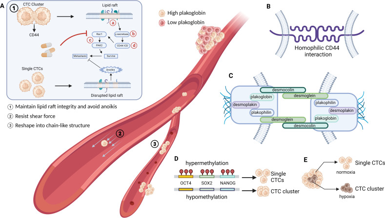
Once clusters can be recovered, the next question is why travelling together benefits tumour-cell survival in the circulation. Figure 2 focuses on homotypic clusters composed of tumour cells. A introduces survival and mechanical advantages, B–C describe the contacts that maintain the group, and D–E connect clustering to epigenetic state and hypoxia.
AThree ways clustering addresses the challenges of circulation
Panel A combines signalling, mechanical protection and shape change in three scenes within the vessel. Together they explain how a cluster can remain viable after leaving the primary tumour and continue moving through the circulation.
① Preserving lipid rafts and resisting anoikis
CD44-positive clusters maintain membrane lipid-raft integrity. The review describes γ-secretase cleavage of CD44, release of its intracellular domain, CD44 ICD, and activation of Rac1–PAK2 survival signalling. The contrasting single CTC has disrupted lipid rafts and is more susceptible to anoikis. Red labels a–d identify potential intervention levels: lipid rafts, γ-secretase, the Rac1–PAK2 pathway and CD44 ICD, respectively.
② Shielding inner cells from shear stress
Cells at the outside of the cluster can protect cells within it from fluid shear. This is a mechanical benefit of collective organisation, complementing the intracellular survival signalling illustrated in ①.
③ Rearranging into a chain to cross narrow vessels
Clusters need not remain compact spheres. They can reorganise into chain-like shapes and traverse capillary-sized channels. Their larger aggregate size therefore does not mean that small vessels must block them; shape plasticity contributes to their ability to disseminate.
BHomophilic CD44 interactions: bringing tumour cells together
CD44 on neighbouring tumour cells can engage in homophilic interactions, establishing contacts that promote homotypic clustering. This panel highlights CD44 at the level of intercellular adhesion.
That is distinct from CD44 cleavage and intracellular signalling in A①: B explains how cells come together, whereas A① explains how the clustered state can support survival. The two levels together motivate the review's discussion of CD44-related intervention strategies.
CPlakoglobin and junctions: maintaining the collective structure
The drawing includes junctional components such as desmoglein, desmocollin, plakoglobin, plakophilin and desmoplakin. The review identifies plakoglobin as important for adherens-junction and desmosome formation, linking these structures to the generation and stability of CTC clusters.
This extends the receptor contact in B: cells require junctional organisation as well as initial attachment to remain a coherent group. The high- and low-plakoglobin states illustrated in the vessel reinforce this connection with clustering.
DHypomethylation and stemness: a molecular state beyond aggregation
Hypomethylation at binding sites for transcription factors including OCT4, SOX2 and NANOG is linked to stemness- and proliferation-associated programmes in CTC clusters. The diagram contrasts hypermethylated sites beside a single CTC with hypomethylated sites beside a cluster.
The discussion draws on single-cell epigenomic profiling. It presents clustering as a molecularly distinct state, not merely physical aggregation; the schematic should not be read as a universal single-switch model in which methylation alone determines every clustering event.
EHypoxia: connecting the primary-tumour environment to clustering
Hypoxia can favour cluster generation by increasing cell–cell junctional components. The normoxia and hypoxia branches connect conditions in the primary tumour with the single-cell or clustered state in which tumour cells enter circulation.
The review discusses breast-cancer mouse models in which changes in primary-tumour burden did not necessarily track changes in CTC clusters or metastasis. The implication is to distinguish primary-tumour effects from effects on clustering and dissemination when studying vascular and hypoxia-related interventions.
Takeaway
Homotypic clusters combine several advantages: adhesion and junctions maintain the group, survival signalling counters anoikis, collective structure and shape change accommodate blood flow, and epigenetic and hypoxic states regulate clustering. Figure 3 then asks what additional functions arise when the travelling partners are blood or stromal cells rather than other tumour cells.
Heterotypic CTC clusters and potential therapeutic targets
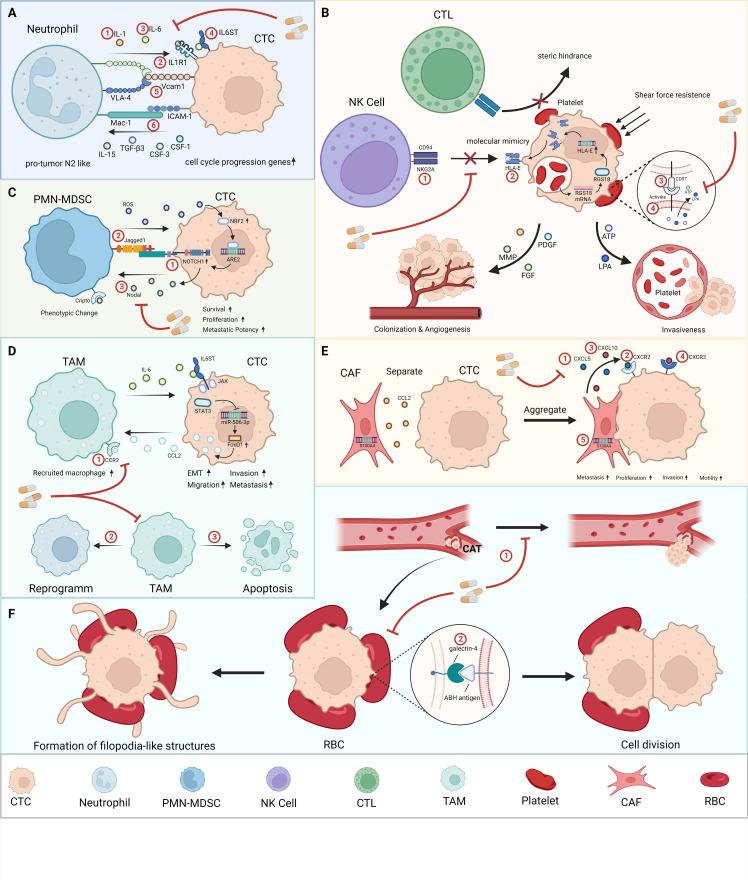
Figure 3 moves from cooperation among tumour cells to cooperation with other cell types. Travelling partners do not all provide the same benefit: some promote proliferation, others shield CTCs from immune attack, and others modify adhesion, matrix or mechanical support. Panels A–F connect these interactions to candidate intervention points; red numbers identify the targets or strategies discussed in each panel.
ACTC–neutrophil: converting adhesive contact into proliferative support
Neutrophils associated with CTCs can provide cytokines such as IL-1β and IL-6 that stimulate cell-cycle progression and proliferation-related gene expression. The review also discusses a reciprocal influence: CTCs within clusters may promote neutrophil activation.
Adhesive interfaces including VCAM1–VLA-4 and MAC-1–ICAM1 help establish contact between the two cell types. The intervention logic has two complementary levels: interrupt IL-1/IL1R1 and IL-6/receptor-related signalling to weaken proliferative support, or interfere with adhesion to affect cluster formation itself.
BCTC–platelet: mechanical shielding, immune escape and vascular effects
Platelets surrounding CTCs can protect them from shear while creating a physical barrier to immune-cell access. They can also transfer MHC-I to tumour cells, providing molecular mimicry. The platelet-derived RGS18–HLA-E mechanism described in the review additionally suppresses NK-cell killing through the HLA-E:CD94–NKG2A axis.
A second branch connects platelet activation to dissemination. Tumour-cell CD97 can activate platelets and promote release of ATP and LPA. The review links ATP to disruption of endothelial junctions and LPA to increased invasiveness; the PDGF, VEGF, MMP and FGF branch extends the discussion to angiogenesis and colonisation.
Candidate approaches include blocking NKG2A or HLA-E and interfering with CD97-related activation. The caption also proposes platelet-membrane-based drug delivery. That is a delivery strategy exploiting platelet properties, rather than another example of directly blocking a receptor.
CCTC–PMN-MDSC: reinforcing paracrine and contact-dependent signals
This panel focuses on polymorphonuclear myeloid-derived suppressor cells, or PMN-MDSCs. Nodal–Cripto paracrine signalling participates in their protumourigenic phenotypic change, followed by ROS-related responses and increased Notch1 expression in CTCs. Contact-dependent Notch1–Jagged1 signalling connects this interaction with enhanced CTC survival, proliferation and metastatic potency.
The mechanism is therefore more than one-way protection by an immune cell: the partners influence both cell state and signalling. The candidate intervention points target the Notch1–Jagged1 axis and Nodal-related communication to interrupt this cooperative, metastasis-promoting interaction.
DCTC–TAM: recruitment, reprogramming and EMT
Tumour-associated macrophages, or TAMs, frequently have M2-like features. CCL2–CCR2 in the figure connects to macrophage recruitment, while signalling between TAMs and CTCs through the JAK2/STAT3/miR-506-3p/FoxQ1 axis is associated with epithelial-to-mesenchymal transition, migration, invasion and metastasis.
The three intervention levels below are interference with CCR2-related recruitment, reprogramming TAMs towards a more antitumour phenotype, and TAM depletion. The review also notes that the mechanisms underlying physical CTC–macrophage clusters require further investigation. The assembled interactions should not all be treated as having been verified within the same circulating cluster.
ECTC–CAF: direct contact, paracrine exchange and matrix support
Cancer-associated fibroblasts, or CAFs, travelling with CTCs can support survival and proliferation through direct contact and paracrine communication. The transition from separated cells to an aggregate highlights that contact itself changes their exchange. The text describes contact-dependent changes involving CXCL10/IP-10 and CCL5/RANTES and discusses the contribution of S100A4-associated fibroblast migration to metastasis.
CAFs can also provide mechanical support against shear and anoikis and modify extracellular-matrix structure to facilitate colonisation. Candidate interventions therefore span chemokine/receptor signalling, including CXCL5- and CXCL10-related pathways, and S100A4, rather than focusing only on breaking a single physical attachment.
FCTC–red blood cell: mechanical attachment, extravasation and division
CTC–red blood cell interactions remain comparatively underexplored. The review proposes that accompanying RBCs may support steady rolling along vessel walls at low flow, increasing opportunities for endothelial adhesion and subsequent extravasation.
Galectin-4-related binding connects RBCs to the tumour-cell surface. The text discusses RBCs as surrogate mechanical attachment surfaces that support adhesion and proliferation; the lower branches depict filopodia-like structures and cell division. The upper branch introduces cancer-associated thrombosis, or CAT. CAT dissociation and galectin-4 interference are presented as potential research directions, not established antimetastatic treatments.
Takeaway
The sequence is partner-cell composition → specific interaction → a survival, immune-evasion or colonisation advantage → a candidate intervention. The review assembles evidence from different cancers and experimental systems; it does not establish that every proposed target clinically eliminates CTC clusters or prevents metastasis. Across all three figures, the logic is to recover and identify intact clusters, understand their cooperative mechanisms, and then investigate interventions matched to their composition.
My contribution
I am the first author of this paper.
Outcomes & record
Published in BMJ Oncology in 2024, volume 3, article e000437. DOI: 10.1136/bmjonc-2024-000437.